Chapter 4 : Motor Control

Brain: Contents Page


An Overview of the Anatomy of Motor Pathways in the CNS


Coronal Section of the Cerebral Hemispheres showing the Internal Capsule

  1. The White Matter is composed of axons whose function is to conduct electrical signals rapidly from one site to another in the CNS.
  2. Gross examination of sections of the brain display white matter
  3. Within each section of the white matter there are thousands if not millions of nerve fibres and gross examination does not provide information about their origin or destination (target).
  4. However gross visual examination identifies certain areas that have important functions.
  5. One such structure is the corpus callosum which carries axons from one hemisphere to the other.
  6. Another is the Internal Capsule, a narrow area where axons communicating between the cortex and the brainstem or spinal cord pass between the caudate nucleus and the putamen, globus pallidus and thalamus
  7. The internal capsule is of particular importance because it is supplied by the middle cerebral artery, which can become occluded and cause stroke.

Artist's Impression of the white matter of the brain (http://johnjchampionseatacarticle.org/)

Five Mysteries of the brain


Top


The Corticospinal Tract

The Main Pathway from the Motor Cortex to the Spinal Cord via the Corona Radiata, Internal Capsule and Brainstem

The motor cortex contains a map of the musculature of the body, and when we perform a voluntary movement, instructions are sent from this area of cortex to the motoneurones using a fast myelinated pathway - the corticospinal tract.

The map of the musculature isn't a simple map of individual muscles, but involves groups of muscles concerned with the same movement. So movements rather than muscles are the nature of the signals emanating from the motor cortex.


*

The Corticospinal Tract

The main motor pathway, responsible for the execution of voluntary movements, begins in the motor cortex and is called the corticospinal tract.

The size of this projection is large enough to be traced by dissection of the brain, and these nerve fibres pass through the corona radiata to the internal capsule, the cerebral peduncle, the pons and medulla.

In the medulla 90% of the fibres cross to the opposite side in the decussation of the pyramids then pass down the lateral columns to reach the motoneurones on the side of the spinal cord opposite the motor cortex (the Lateral Corticospinal Tract).

It is because of this decussation in the pyramids of the medulla that a lesion in the internal capsule causes paralysis on the opposite side of the body.

Another name for the corticospinal tract is the Pyramidal Tract (because the axons pass through the pyramids).

A smaller percentage of the descending fibres form the anterior corticospinal tract within the spinal cord, and pass down the cord ipsilaterally before crossing the midline at a segmental level to innervate the contralateral motoneurones.

*

90% of corticospinal tract axons follow the path shown above.


More Details on the Anatomy of the Cortico-Spinal Tract

Corona Radiata
Internal Capsule
Cerebral Peduncles
Pyramids of the Medulla
Lateral and Anterior Columns of the Spinal Cord


http://www.docstoc.com/docs/91260976/Cerebellum-Basal-Ganglia-Motor-Cortex
www.utdallas.edu

Top


Cortico-Bulbar Pathways and the Control of Facial Muscles

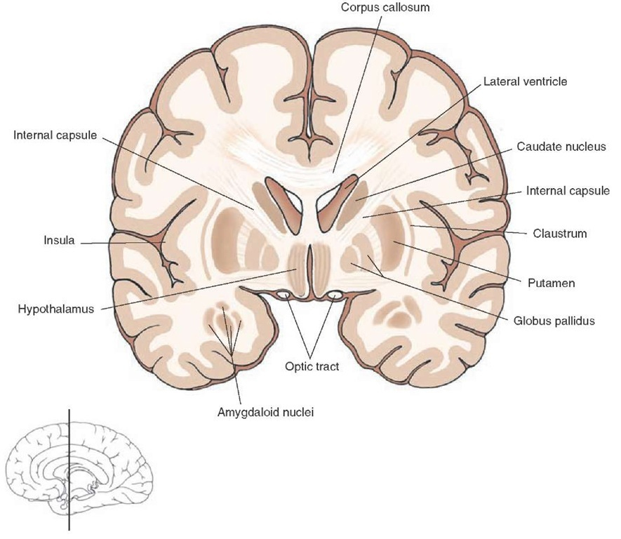
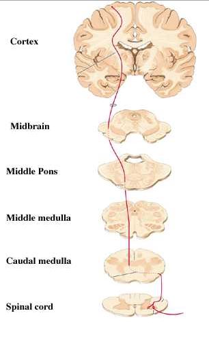
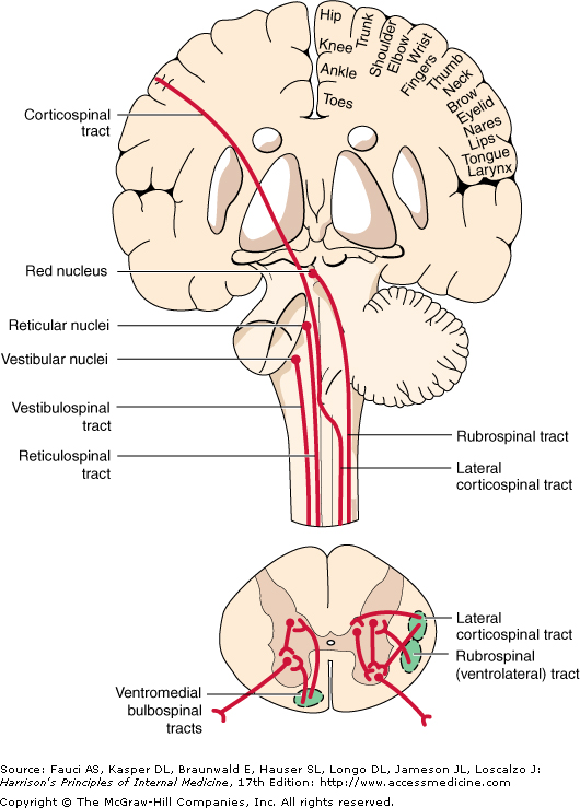
The cortex communicates with the brainstem using several types of cortico-bulbar axons that terminate in:

  • motor nuclei of the cranial nerves
  • pontine nuclei that project to the cerebellum
  • the red nucleus of the midbrain, the origin of the rubro-spinal tract
  • the brainstem reticular formation, the origin of the reticulo-spinal pathways

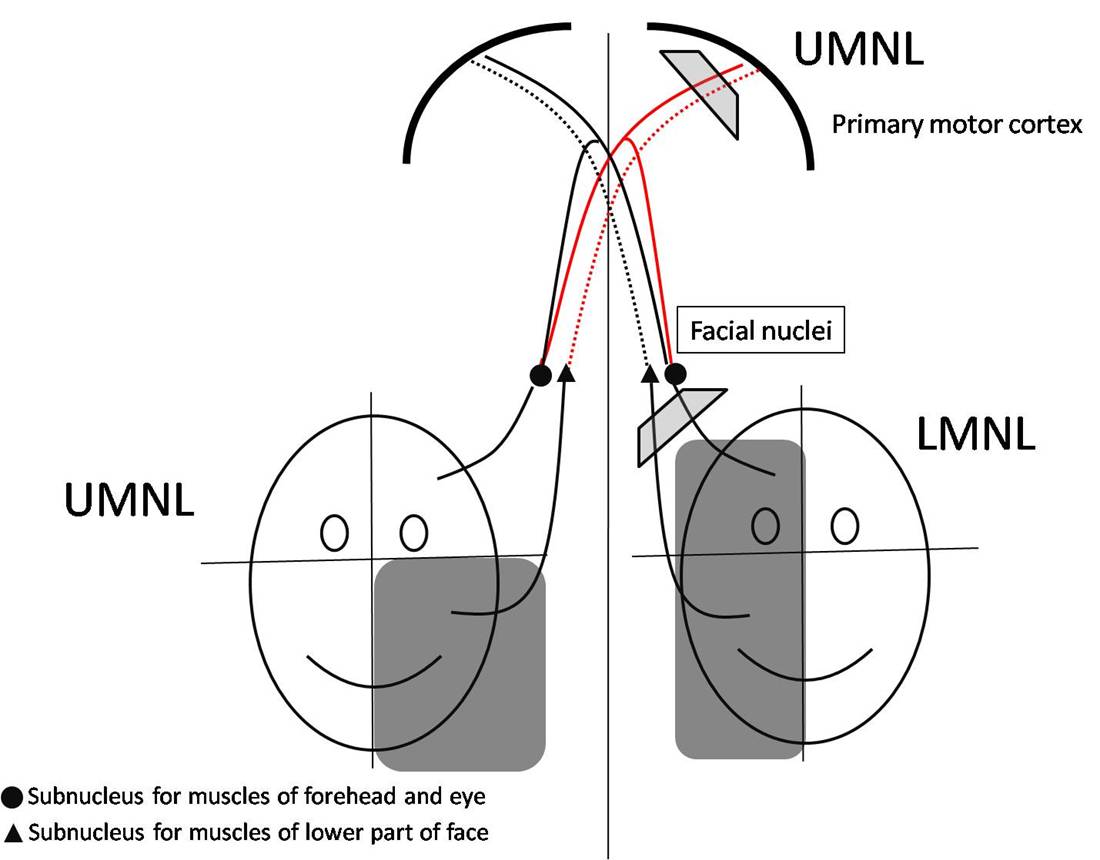
Pathways from the face and neck area of the motor cortex project to motoneurones in the brainstem (hence the term cortico-bulbar tract). They innervate motor nuclei including the facial and trigeminal nerve nuclei, that are concerned with the muscles of facial expression and mastication respectively; and muscles in the tongue and neck.

For muscle groups below the eyes, the cortico-bulbar tracts are crossed, so one side of the brain controls the muscles on the opposite side of the face and neck. The cranial nerves involved are the accessory and hypoglossal nerves, innervating the muscles of the neck and tongue.

However, for the upper part of the face, innervated by the facial nerve, the motoneurones are innervated from both sides of the cortex; this involves the part of the facial nucleus innervating the forehead and eyelids. In contrast the part of the facial nerve nucleus that innervates the lower part of the face- cheek and lips - are innervated only from the contralateral cortex.

Significantly the eyebrows can be raised on both sides of the face in many patients with a stroke (because of the bilateral innervation of these motoneurones), unlike the muscles of the lower part of the face, which are paralysed on the side opposite the cerebral lesion.

In contrast, a lower motoneurone lesion to the facial nerve however paralyses all muscles on one side of the face (as in Bell's Palsy).

*
www.webmedcentral.com

Top

Other Pathways involved in Motor Control: Bulbo-Spinal Pathways
*

Details of Bulbo-Spinal Pathways
  • Reticulo-spinal tracts
  • Rubrospinal tract
  • Vestibulo-spinal tracts

Harrison's Internal Medicine McGraw Hill

Top

Clinical Anatomy

Stroke Stroke is a common neurological disorder that develops as a result of occlusion of arteries supplying important areas of the brain; the most common strokes are the result of occlusion of the middle cerebral artery. There are three main types of vascular pathology:

Embolism: a clot of blood forms in the heart (often associated with atrial fibrillation) and the clot enters the aorta and travels to the middle cerebral artery, blocking it and depriving the neurone of their blood supply

Thrombosis: changes in the interior of the middle cerebral artery cause occlusion of the blood supply

Haemorrhage: the middle cerebral artery is weakened and may burst releasing blood into the internal capsule and other structures.

Top

Chapter 4 : Motor Control

Brain: Contents Page

© HumanPhysiology.Academy 2014-2015